 | |||||||||||||||
 |
 |
 |
 |
 |
 |
 |
 |
 |
 |
 |
 |
 |
 |
 |
 |
V-CEM
VANDERBILT
CRYO
ELECTRON
MICROSCOPY
V-CEM Only:
Cryo TEM
Single partilce
This method is used to obtain high resolution 3D structure of protein complexes.
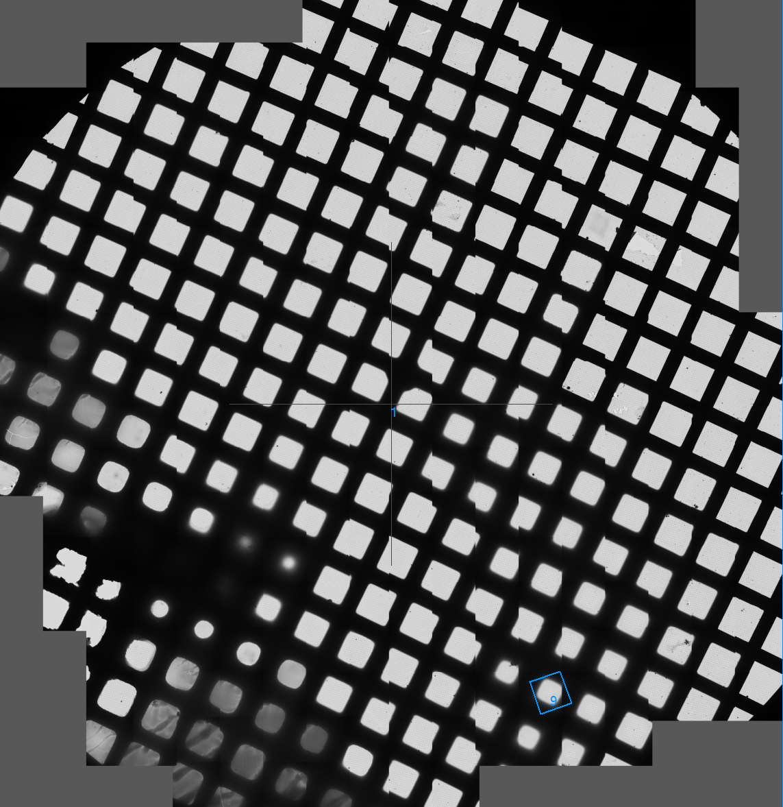
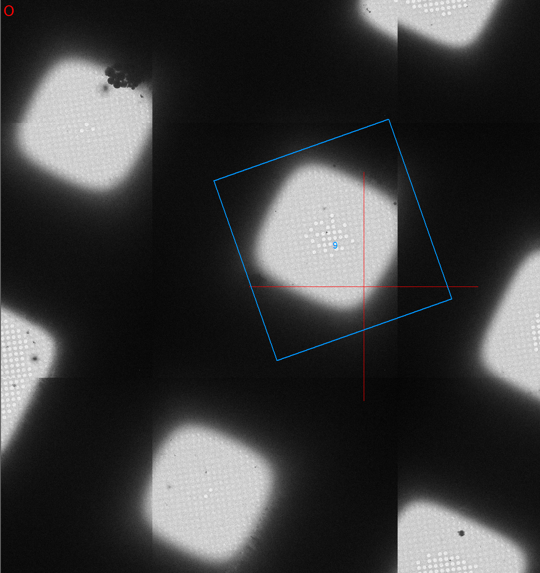
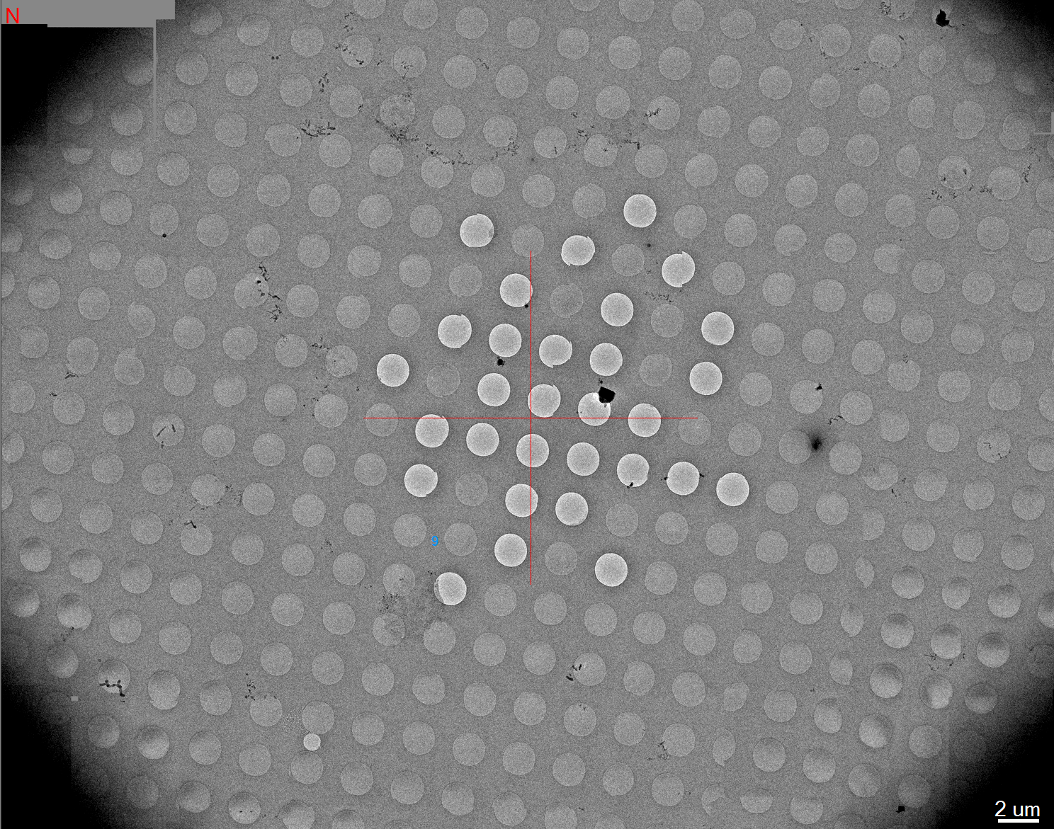
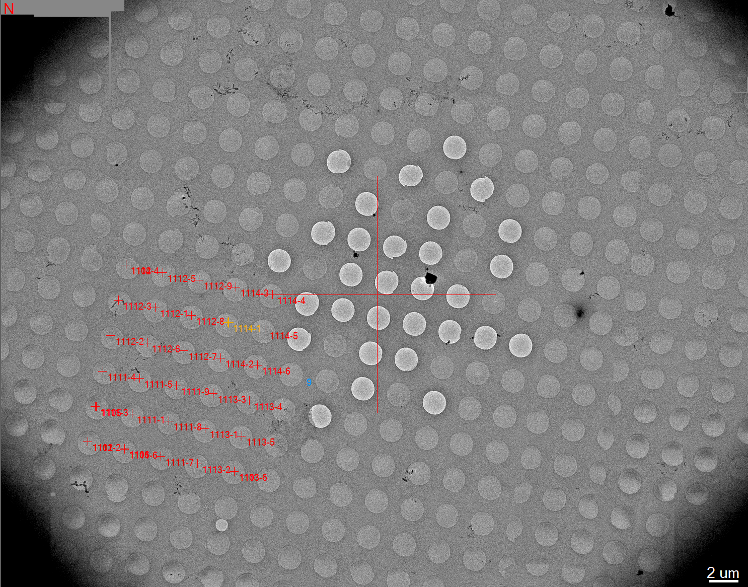
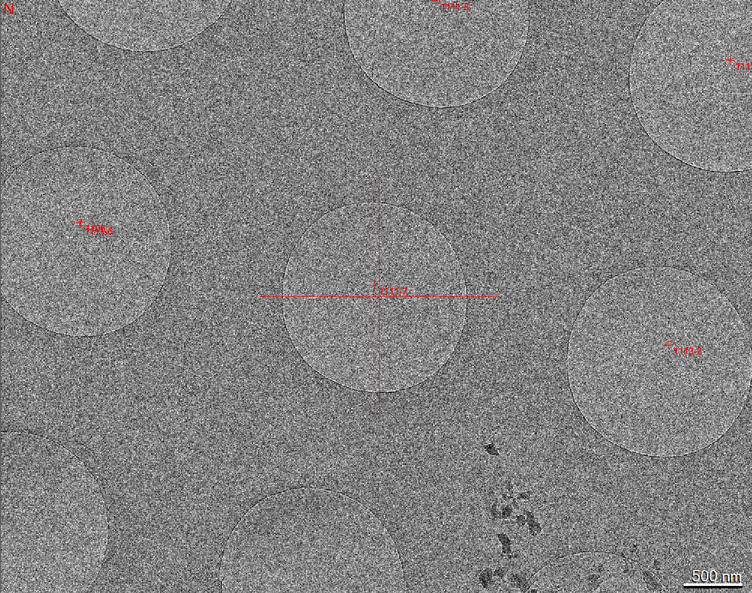
The sample (1-3 ul) applied to holey carbon grid and plnuge frozen in layer of vitreous ice that preserved the netive condition. The sample image at cryogenic temperatures by direct electron detectors (K2-Polara) or CCD (F20). The low contrast of this method limit the size of the biological sample to above ~200kDa.
The direct electron detectors increase the image contrast and with movie mode (a series of low dose images) allowed drift correction.
The data collection is done semi-automated with SerialEM and the micrographs are preprocess on-the-fly by GPU computer to get sense of the quality of the data.
|
VU Home |
VUMC Home |
People Finder |
University Calendar
webmaster- modified on March 14, 2017 |
